ECG Interpretation Made Easy (Learn How to Interpret an ECG in 13 Minutes)
3-Second Blueprint (English)
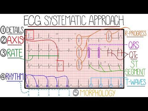
- Confirm 25 mm/s and 10 mm/mV calibration, then map 12 leads across lateral, inferior, and septal-anterior cardiac territories.
- Evaluate axis, rate (60–100 bpm), RR-interval regularity, and P-wave-to-QRS pairing to identify arrhythmias and heart blocks.
- Check for pathological Q waves and ST-segment elevation with reciprocal depression, always correlating findings with history.
12-Lead ECG TerritoriesCalibration (25 mm/s, 10 mm/mV)Rate & RR-Interval RhythmPathological Q WavesST Elevation & Reciprocal Change
3-Sekunden-Fazit (Deutsch)
- Kalibrierung (25 mm/s, 10 mm/mV) prüfen und die 12 Ableitungen lateralen, inferioren sowie septalen-anterioren Arealen zuordnen.
- Lagetyp, Frequenz (60–100/min), RR-Regelmäßigkeit sowie P-Wellen-QRS-Überleitung auf Arrhythmien und AV-Blöcke analysieren.
- Pathologische Q-Zacken sowie ST-Hebungen mit reziproken Senkungen prüfen und stets mit der Klinik des Patienten abgleichen.
12-Kanal-EKG-WandarealeKalibrierung (25 mm/s, 10 mm/mV)Frequenz & RR-RhythmusanalysePathologische Q-ZackenST-Strecken-Diagnostik
Note / Hinweis: Blueprints are AI-generated educational summaries (0% Politics · 0% Religion · 0% Brainrot) and do not constitute medical, legal, or financial advice. / KI-generierte Zusammenfassung zu Bildungszwecken – ersetzt keine medizinische, rechtliche oder finanzielle Fachberatung.